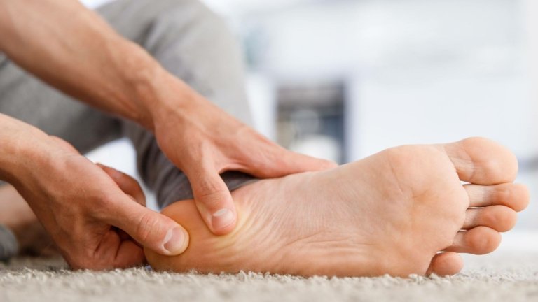
A heel spur is a term for a growth on the heel that is present in the form of a calcium deposit or bone spur or osteophyte. Heel spurs have an overall prevalence of 15%, and are more common in the elderly, with 35% of individuals in the 60–69 age group having heel spurs. A heel spur can also be asymptomatic, meaning it does not cause any pain, but it always needs to be treated with physiotherapy as it represents a risk factor for the onset of pain. A heel spur usually hurts on one side, although two thirds of patients also have a spur on the other heel, which they refer to as a problem with the other heel in the past.
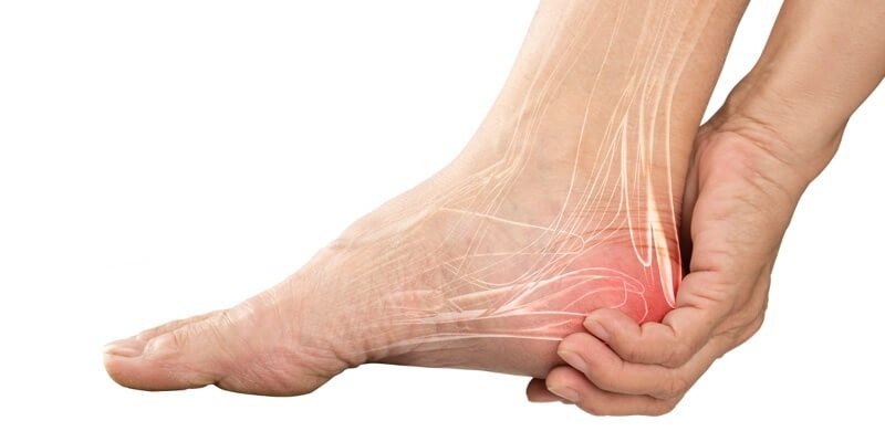
A heel spur can grow up to 1 cm in size and can occur on the back of the heel (spina calcanei superior) as a result of overload of the Achilles tendon attachment point, or on the underside of the heel (spina calcanei inferior) as a result of overload of the plantar fascia attachment point. A plantar heel spur, on the underside of the heel, is a result of plantar fasciitis, and is the more common and commonly referred to “heel spur” by patients upon visit.
Risk factors for developing a heel spur include flat feet, being overweight, wearing high heels, sports overload, especially jumping and running, diabetes, and age. It occurs more often in women, athletes, elderly people between 60 and 69 years of age, and diabetics.
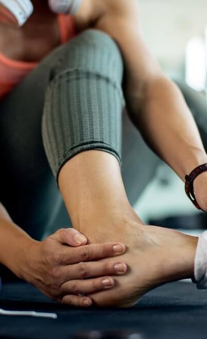
A heel spur has a characteristic pattern of symptoms, making it easy to distinguish from other pathological conditions that cause pain in the area of the calcaneus. The pain of a heel spur is sharp and intense, occurring mainly in the morning with the first steps, in the area of the calcaneus. A characteristic of heel spurs is that the pain disappears or decreases after walking a short distance. Heel spurs are characterised by post-static dyskinesia, meaning that after physical inactivity or rest, the heel always hurts intensely for the first few steps.
The symptoms of a heel spur depend on the size, shape, and location of the spur, but in most cases, the pain is strongest in the morning, as the plantar fascia shortens and expands at night. Symptoms also typically worsen when running and walking.
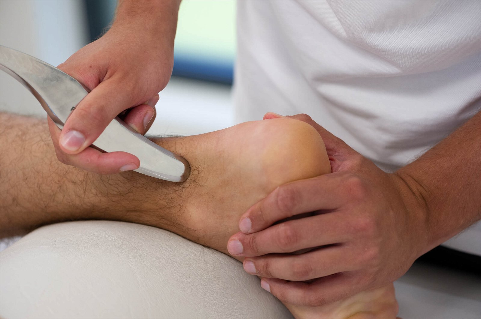
Heel spur is one of the pathological conditions that require a comprehensive conservative approach of physiotherapy and kinesiology. This means that if we only perform one type of therapy in isolation, we will not eliminate the heel spur. Comprehensive treatment includes extracorporeal shockwaves, TECAR therapy, laser therapy, manual therapy, and kinesiology training.
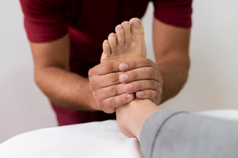
Treatment for a heel spur usually lasts between 16 and 24 weeks; however, if the heel spur has been present for more than a year, treatment is extended. To treat heel spurs, extracorporeal shockwave therapy is often performed in isolation, as it is the most effective method of “breaking up” or eliminating heel spurs, but it must be performed together with special exercises and manual therapy.
Treatment of heel spurs must include techniques to improve the quality of the connective tissue of the plantar fascia, which is achieved through exercises to strengthen the intrinsic muscles of the foot and endurance exercises for shin muscle strength.
At MEDICOFIT clinic, we use EMS focused shockwaves as the leading method of treating heel spurs, which are the most effective tool for heel spur rehabilitation in clinical practice – patients usually notice pain-relieving effects after their first therapy. Extracorporeal shockwaves break down the deposited calcium into smaller units, which are then absorbed and broken down in the surrounding tissue. Extracorporeal shockwave therapy must be performed in combination with special exercises to strengthen the arch of the foot, which we always consider at MEDICOFIT clinic.