How common is osteoarthritis of the knee?
Osteoarthrosis of the knee (OA of the knee) is the most common joint disease in the world. It mostly occurs on joints that carry a lot of weight. Most frequently afflicted is the knee; however, it often also occurs on the hips (3).
What is osteoarthrosis of the knee?
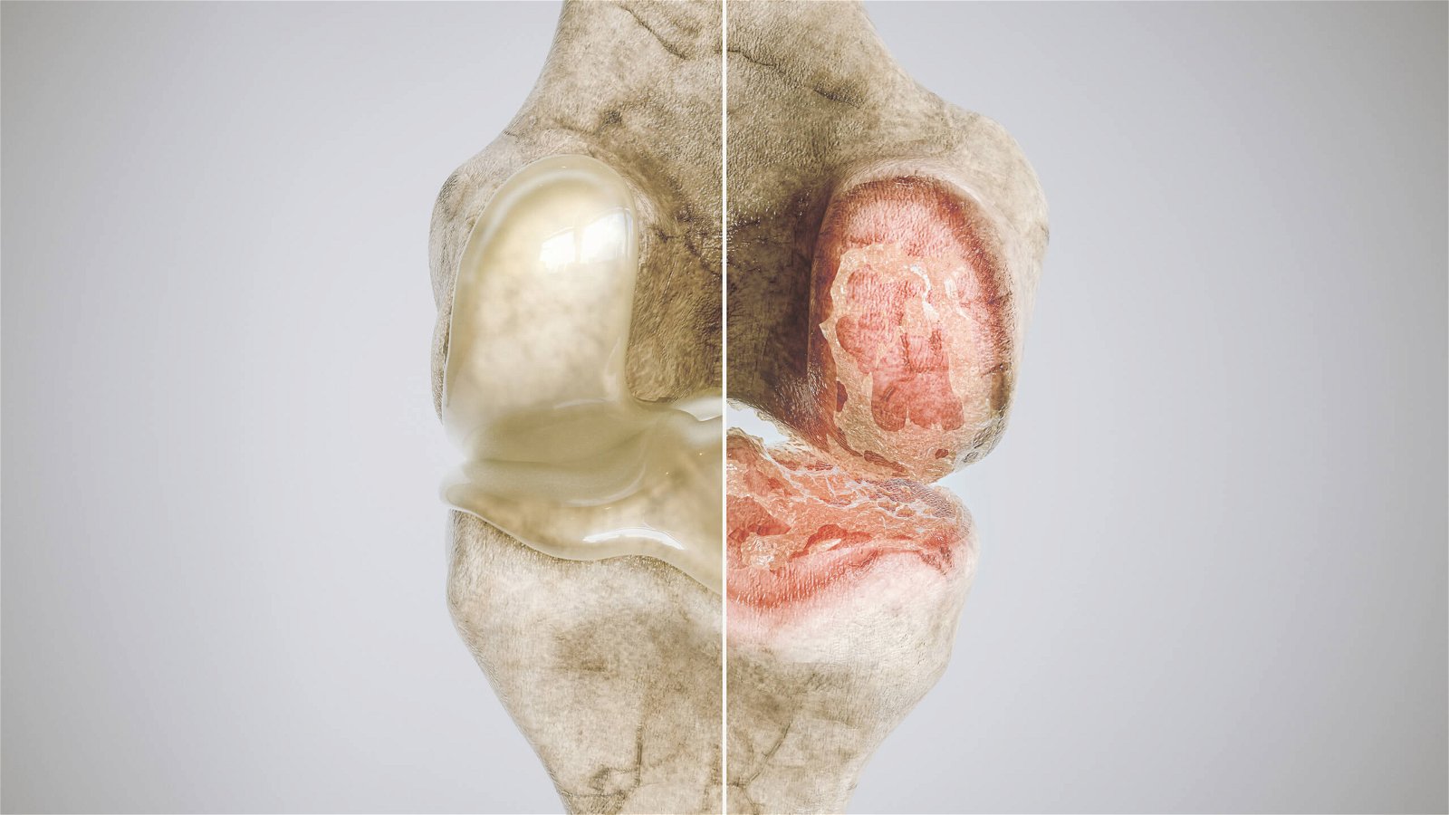
Osteoarthrosis of the knee is a degenerative disease of the synovial joints, the main characteristic of which is premature, progressive deterioration of joint cartilage. Later, inflammation (arthritis) may occur, which is followed by secondary changes that appear on articular and periarticular structures such as the synovial membrane, fibrous membrane, subchondral bone, tendons and muscles (13). The consequent inflammation can also be called osteoarthritis (3).
Which are the typical symptoms and signs of osteoarthrosis of the knee?
Clinically the disease is manifested by pain and loss of joint function. But not all that show radiographic signs of osteoarthrosis of the knee are symptomatic. The radiologic imaging of approximately half of the adult population aged over 60, show signs of osteoarthrosis, but only 15% of these individuals were symptomatic, so experiencing pain and limited mobility (7).
Later on, these symptoms can be joined by crunching or creaking noises in the joint (crepitation), joint effusion due to secondary inflammation, muscle weakness, contracture, ankylosis, and a thickened or deformed joint (13). Pain develops gradually, is profound, dull and causes discomfort. In the initial stage, it occurs after physical activity, while in the advanced stage of the disease it occurs also during the resting period. In the final stage of the disease, the only therapeutic solution is the introduction of the total endoprosthesis of the knee (13).
In 2018 in Slovenia, 90.4 % of all orthopaedic surgeries of the knee were because of the diagnosis of osteoarthrosis (6). It is to be expected that the incidence of osteoarthrosis of the knee condition will continue increasing due to demographic ageing and the obesity epidemic (7, 13).
Osteoarthrosis of the knee is divided into primary and secondary.
Primary osteoarthrosis develops solely due to ageing and is not the consequence of any pre-existing illness or injury (11). It is characterised by genetic predisposition and influence factors such as female sex and obesity (13).
Secondary osteoarthritis, on the other hand, develops from already known pre-existing illness of the locomotive apparatus in the sense of developmental anomalies, injuries, poor joint alignment due to a previous surgery such as meniscectomy, inflammatory changes, metabolic disease, avascular necrosis or excessive long-term strain (13, 3).
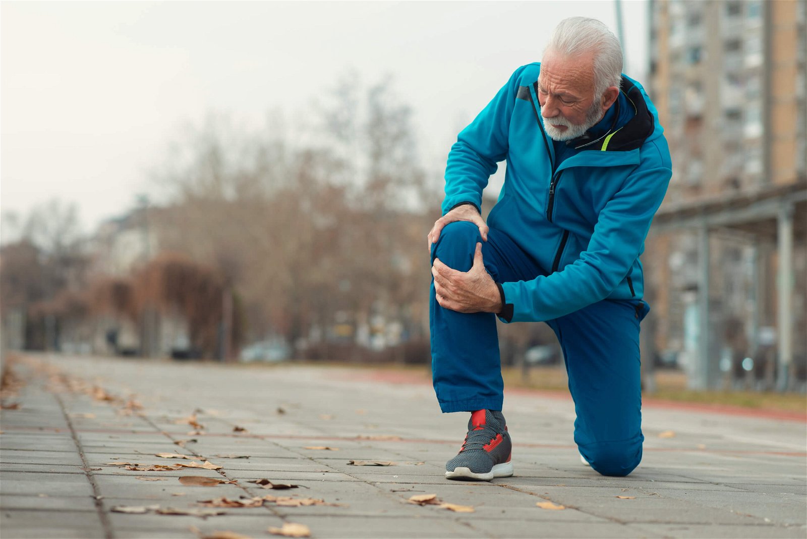
Dividing the line between both forms of osteoarthrosis is difficult in clinical practice as osteoarthrosis is connected to multifactorial etiologic factors and has lately no longer been considered an inevitable part of growing old. Instead, it is closely connected to systemic metabolism (the development of osteoarthrosis is connected to hypertension, hypercholesterolemia, diabetes, inflammation and angiogenesis) (1).
Stages of osteoarthrosis
The development of osteoarthrosis can be described in five phases (3):
1. Disruption of the joint surface
Degenerativna osteoartroza se začne s propadom sklepne površine (3). To se zgodi kot posledica neravnovesja razgradnje hrustanca in njegove sinteze. Vzroki so lahko mehanski (udarci ali torzijske obremenitve na sklep), vnetni (pospešena razgradnja molekul hrustančnega matriksa), metabolni (zmanjšana sposobnost hondrocitov), idr (13).
Normalno gladka sklepna površina se začne lomiti skupaj s kolagenskimi vlakni in površina postane hrapava. Frikcija ob hrapavo površino povzroči, da na površini hrustanca nastajajo delci, ki se absorbirajo v sinovijo, kjer povročajo vnetni odziv. To pa posameznik občuti kot togost in bolečino v sklepu, ponavadi po aktivnosti in ne med njo (3).
2. Irritation of the synovia
The synovia irritation causes the release of various enzymes and cellular processes, which leads to inflammation (3). The synovial capsule and synovial cells play an important role in the processes of joints. The capsule along with the ligaments ensures stability and determines the range of motion. Because these structures are subject to knots and fibrosis during osteoarthrosis, the range of motion is very limited.
3. Transformation of tissue
In the advanced stage of osteoarthrosis, the degenerative process expands deeper into cartilage structure, all the way to the calcined layer or subchondral bone. Cartilage becomes thinner, its volume decreases, and the surface becomes irregular with numerous cracks around which chondrocytes are arranged. Usually, cartilage tissue is replaced by fibrotic tissue (13). Because of the change in the shape and congruity, the weight-bearing pattern changes, which means that other areas of the joint take on the strain (3).
4. Osteosclerosis
With the progression of the disease, the defect can occur on the whole thickness of the cartilage so that the bone is completely exposed (13) and the rubbing of bone against bone is painful. Friction increases and the transfer of weight through the joint becomes uneven. These changes lead, due to overload, to microfractures of bones that are treated with callus, which means that the bone gradually grows thicker, more sclerotic and less resistant (3).
5. Disorganisation
The loss of normal bone structure causes once again various injuries and pain. The joint becomes more rigid and due to bone shortening, the periarticular ligaments and connective tissue also loosen which leads to even bigger joint instability (3).
Path to diagnosing the osteoarthrosis condition
The path to correct diagnosis includes a physical examination, clinical joint testing by a physiotherapist and referral for x-ray imaging and/or an MRI. In some cases, they might carry out an exploratory arthroscopy, where, during the operation, they insert a camera, with which they examine the damaged tissue (10).
Knee osteoarthritis can be divided into 5 stages (5,10) depending on the degree of impairment:
Stage 0: represents a healthy, functional knee.
Stage 1: minimal pathological changes are present, but no pain or discomfort is present.
Stage 2: symptoms such as joint pain after prolonged physical activity appear. X-ray imaging can already show several pathological changes, but at this stage the cartilage is still mostly preserved.
Stage 3: pain in this joint occurs more often, especially after prolonged periods of sitting. Noticeable changes are visible in the cartilage, and the joint space is getting smaller.
Stage 4: represents the highest number of osteoarthritis. At this stage, the joint space is dramatically reduced and the cartilage tissue is almost gone. The content of synovial fluid is increased. Individuals in this phase cause high pain that does not subside even during rest.
Physical Examination of the Knee
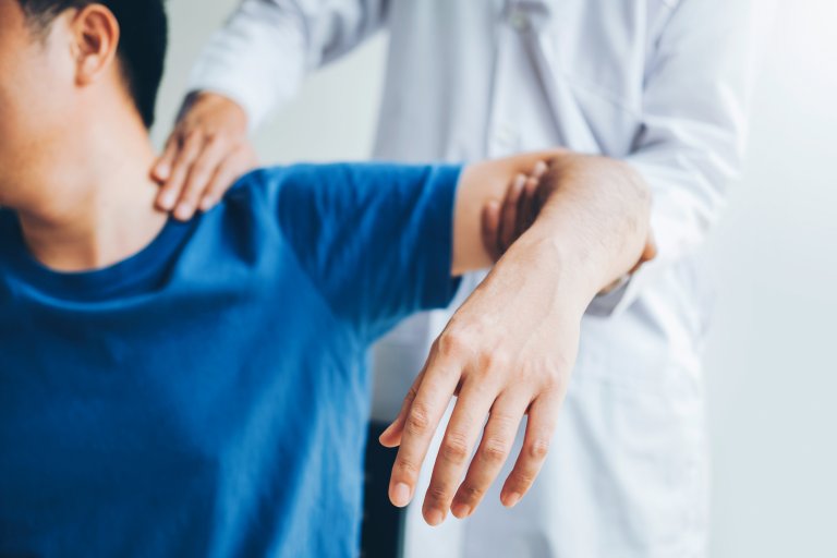
The most important part of diagnosing the condition of osteoarthritis is the clinical examination, which by the physiotherapist includes: visual assessment of the injured knee: the knee may be swollen and red; observation of functional patterns of movement and balance: the physiotherapist will examine your gait and assess whether you need technical aids such as crutches or a stilt.
He/she will examine your movement patterns when getting up or sitting down on a chair, walking up the stairs, etc.; by feeling the injured knee, the physiotherapist will assess the swelling, the change in skin temperature and the sensitivity of the area; joint mobility measurements in passive and active mode will be required. This assesses the range of motion you achieve between knee flexion and extension.
The muscle status is also assessed, in which a muscle deficit is expected on the knee with a state of osteoarthrosis (osteoarthritis?) (10).

Treatment of Osteoarthritis of the Knee
Osteoarthritis of the knee is the most common diagnosis for total knee endoprosthesis. However, since the knee joint is the most complex joint in the human body, from a biomechanical point of view, the insertion of a knee endoprosthesis is complicated. Despite modern techniques, implants, improved pre- and post-operative rehabilitation protocols, it is still not possible to achieve a completely “normal” knee with the insertion of an artificial joint. (9).
Therefore, it is extremely important to completely exhaust conservative treatment before surgery, which includes special physiotherapy and exercise protocols. With high-quality rehabilitation we will extend the time until surgery for osteoarthritis of the knee, if only this is imminent, and at the same time reduce pain and improve the strength of the lower extremities, the range of motion and the performance of functional tasks. This will positively contribute to a better quality of life despite the potentially advanced state of knee osteoarthritis(2).
According to the latest clinical guidelines (2020), the positive effects of physiotherapy and kinesiology exercise outweigh the potential side effects and worsening of symptoms.
These were rarely reported. The guidelines therefore advise that regular and directed physiotherapy and special physical exercise should be included in the daily life of an individual with knee osteoarthritis (12).
The importance of exercising
It is important to start each exercise unit with a warm-up and end it with a cool-down. The general goals in the exercise process and physiotherapy for an individual with knee osteoarthritis are to reduce pain and inflammation in the knee joint, normalize range of motion, improve muscle strength of the lower extremities, improve proprioception, agility and balance, and generally improve physical condition (10). Physiotherapy with modern diamagnetic therapy and the application of a therapeutic laser is very effective.
- Frequency of Training Units
For the conservative treatment of osteoarthritis of the knee, daily physical activity is recommended or at least 5 days a week for 30 minutes of special strength training. Targeted physiotherapy with a specialist in degenerative conditions is recommended at least 2 times a week.
- Physiotherapy and Special Exercise in the Condition of Osteoarthritis of the Knee – Intensity
When training for the acquisition of special muscle strength, in order to reach the minimum standards, it is necessary to perform 8–15 repetitions with 2–4 sets of 60–80 % 1RM (1 repetition maximum) (values 14–17 according to the Borg effort scale) or 50–60 % 1RM (12-13 Borg scale) for individuals who are beginners in strength training (12). The minimum standard must be performed for special exercises of the open kinetic chain, for which we have prepared protocols in the Medicofit clinic for the condition of osteoarthritis of the knee. Physiotherapy must first apply isometric exercises for the condition of osteoarthritis of the knee to prepare the muscle groups for later more intensive loading.
To gain aerobic endurance, guidelines state that the level of rehabilitation exercise should be adjusted to > 60 % of maximum heart rate (14–17 Borg scale) or 40–60 % of maximum heart rate (Borg scale 12–13) for beginners (12). Physiotherapy must always take place simultaneously with kinesiological treatment in the case of knee osteoarthritis.
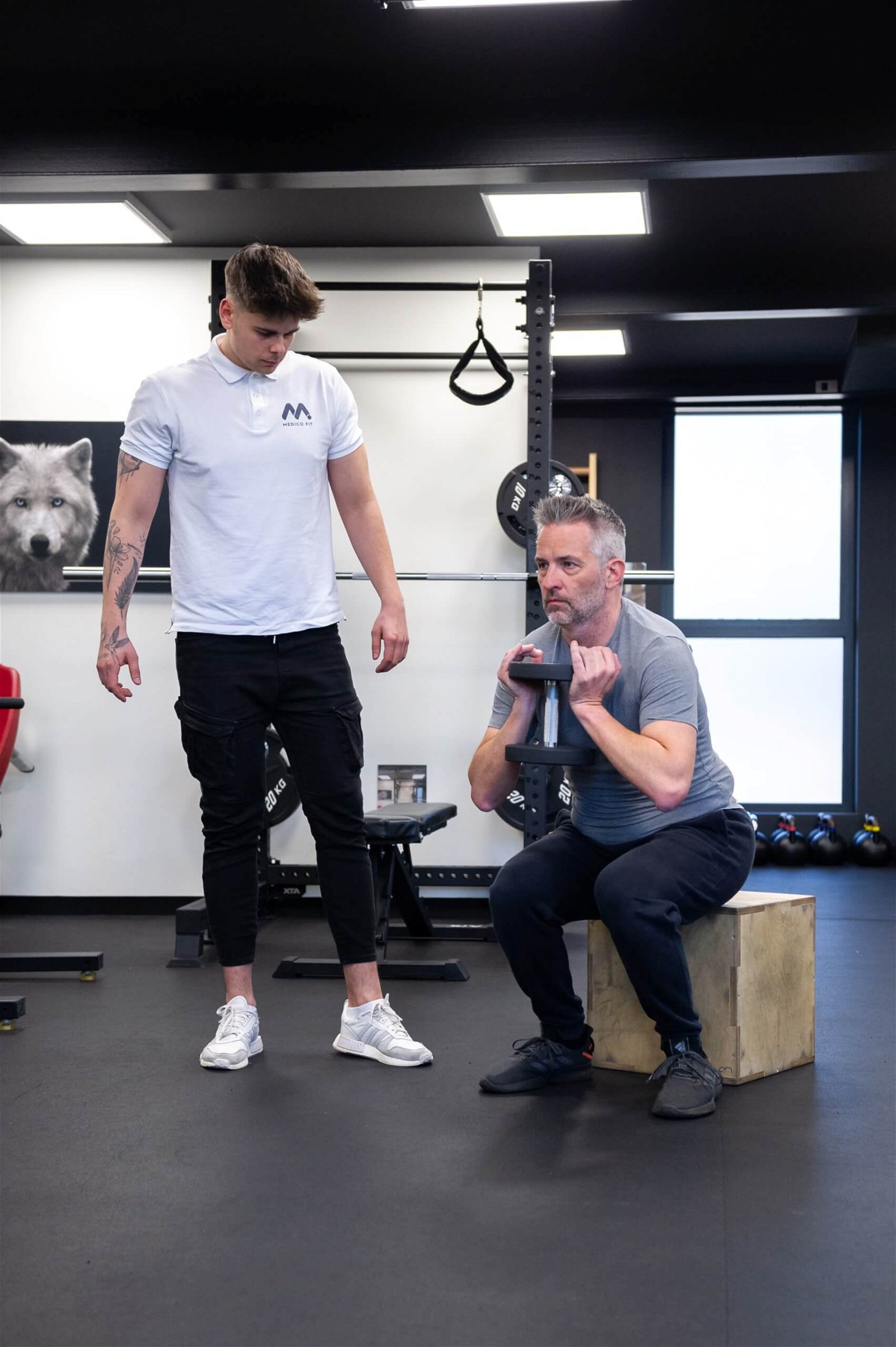
A qualified physiotherapist who will accompany you in the rehabilitation process will determine the initial intensity of strength training and monitor it during the training process and increase it appropriately over time. The intensity of strength training and aerobic training is checked once a week and is increased to the highest level according to the individual’s ability. If joint pain occurs after a training session and does not go away after more than two hours, the next training session should be reduced in intensity (12). A well-qualified kinesiologist or physiotherapist will use variations in sets and repetitions, duration of the set or exercise, type of exercise and rest to adjust the intensity, and in this process will actively cooperate with you and your subjective perception as well as your well-being. Physiotherapy is intensive especially in the initial phase of treatment, it aims to reduce active symptoms in order to prepare the knee joint for the application of strength exercises.
- Type of Exercise, Physiotherapy and Kinesiology in Osteoarthritis of the Knee
When exercising muscle strength for osteoarthritis of the knee, it is necessary to choose exercises aimed mainly at large muscle groups of the knee and hip muscles. Exercises are performed on both legs even if osteoarthritis of the knee occurs only on one side. Exercises with your own weight and resistance exercises using crosstrainers or free weights can be performed. Activities with a relatively low load on the joints, such as walking, cycling, swimming or rowing, are chosen for aerobic exercise. If the problem is maintaining balance, it is necessary to include specific tasks for increasing balance and neuromuscular coordination in training units. Also, depending on individual needs, specific exercises are added to increase the range of motion if there is muscle shortening or reversible limitations of joint mobility (12).
- Duration of Treatment of Osteoarthritis of the Knee, Physiotherapy
There is no cure for osteoarthritis of the knee, and once the disease has developed, it is necessary to try to prevent the progression of the disease (10). We recommend at least a 3–4 month cycle of physiotherapy treatments with check-ups after the end of this period, e.g. after 3 and 6 months. Self-exercise — even in the home environment — is encouraged with check-ups (12). At the Medicofit clinic, we provide comprehensive treatment in check-ups in the diagnostic physiotherapy department. Proper and regular exercise at home brings good results and improves physical condition, and it has been proven that individuals who take part in specially guided exercise units within the framework of physiotherapy and kinesiology resort to analgesics less often and are more satisfied with the rehabilitation process (4).
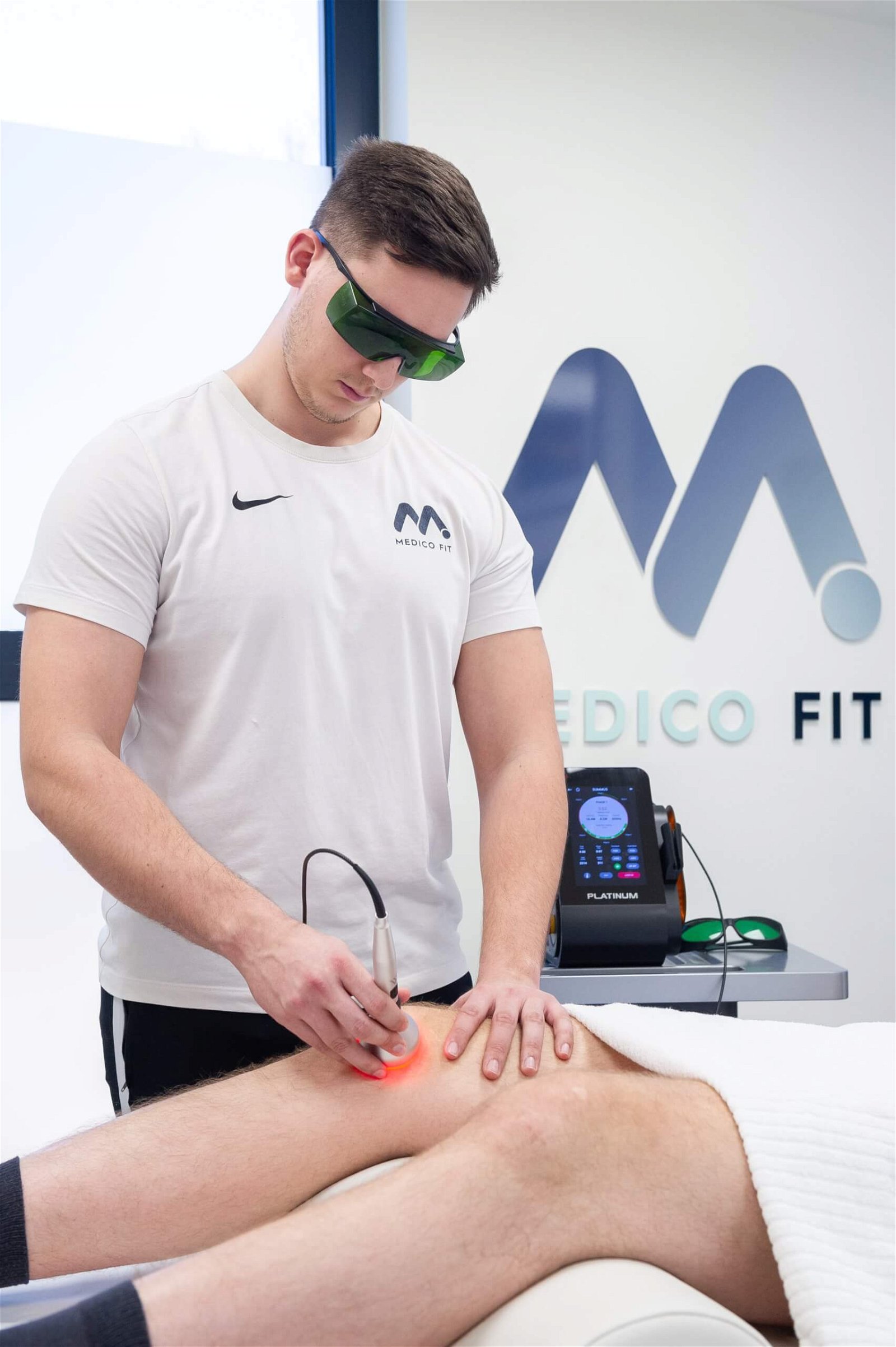
As experts in the field of physiotherapy and/or kinesiology, who understand the pathological background of the condition of osteoarthritis of the knee, will be able to choose, together with you, the appropriate difficulty of the special exercise with which you will make good progress. He will also be able to recognize so called red flags, which are conditions in which it is necessary to stop exercising or adapt it, adjust physiotherapy techniques and consult with the doctor about the further course of rehabilitation.
Red flags for osteoarthritis of the knee are a warm and swollen (red) knee, unexplained severe knee pain, a locked joint, severe pain at rest without traumatic injury, increased pain that does not subside with analgesics, inability to bear weight on the leg if before this was possible, the occurrence of pain in the swords during dorsiflexion of the toes, and the development of fever > 38.5 °C (12). At the Medicofit clinic, we are specialists in the treatment of acute symptoms and red flags of osteoarthritis of the knee. Common mistakes in the rehabilitation process also occur because individuals with osteoarthritis of the knee tend to strengthen the knee muscles, but neglect to strengthen the hip muscles.
Physiotherapy must be specially individually adapted to the patient’s pathology. When evaluated, patients with osteoarthritis of the knee, according to studies, often have weakness of the hip muscles and are consequently more inclined to strain the medial part of the knee joint, which leads to an additional worsening of the already existing disease of osteoarthritis of the knee. Hip strengthening exercises have been shown to improve lower extremity mechanics and reduce knee strain (9).
- Berger, M., 2002. Novejša spoznanja na področju etiopatogeneze in zdravljenja primarne osteoartroze. Zdrav Vest, 71, pp. 235-9.
- Calatayud, J., Casana, J., Ezzatvar, Y., Jakobsen, M.D., Sundstrup, E. & Andersen, L.L., 2017. High-intensity preoperative training improves physical and functional recovery in the early post-operative periods after total knee arthroplasty: a randomized controlled trial. Knee Surg Sports Traumatol Arthrosc, 25, pp. 2864–2872.
- Dandj, D.J. & Edwards, D.J., 2009. Essential Orthopaedics and Trauma. London: Churchill Livingstone Elsevier.
- Deyle, G.D., Allison, S.C., Matekel, R.L., Ryder, M.G., Stang. J.M., Gohdes, D.D., et al., 2005. Physical therapy treatment effectiveness for osteoarthritis of the knee: a randomized comparison of supervised clinical exercise and manual therapy procedures versus a home exercise program. Phys Ther, 85(12), pp. 1301-17.
- Lespasio, M.J., Piuzzi, N.S., Husni, M.E., Muschler, G.F., Guarino, A.J. & Mont, M.A., 2017. Knee osteoarthritis: a primer. The Permanente Journal.
- Levašič, V., Savarin, D. &Milošev, I., 2020. Valdoltra knee arthroplasty registry report 2002-2019. Valdoltra Arthroplasty registry.
- Michael, J.W., Schlüter-Brust, K.U. & Eysel, P., 2010. The epidemiology, etiology, diagnosis, and treatment of osteoarthritis of the knee. Deutsches Arzteblatt International, 107(9).
- Moličnik, A. & Merc, M., 2010. Endoprotetika kolenskega sklepa. In: VI. Mariborsko ortopedstko srečanje. Interdisciplinarno strokovno srečanje in učne delavnice. Artroza in endoprotetika sklepov. Zbornik vabljenih predavanj. Maribor, pp. 81-93.
- Neelapala, Y.R., Bhagat & M., Shah, P., 2020. Hip Muscle Strengthening for Knee Osteoarthritis: A Systematic Review of Literature. Journal of geriatric physical therapy, 1;43(2), pp. 89-98.
- Physiopedia. Knee Oseoarthritis. Available at: https://www.physio-pedia.com/Knee_Osteoarthritis#cite_note-Krucik-10 [10.9.2021]
- Spinalogy Clinic. Understanding the difference between primary and secondary osteoarthritis. Available at: https://spinalogy.com/understanding-difference-primary-secondary-osteoarthritis/ [10.9.2021]
- Van Doormaal, M.C.M., Meerhoff, G.A., Vliet Vlieland, T.P.M., & Wilfred, F.P., 2020. A clinical practice guideline for physical therapy in patients with hip or knee osteoarthritis. Musculoskeletal Care, 18 (4), pp. 575-595.
- Vogrin, M. & Naranđa, J., 2010. Osteoartroza: Epidemiologija, patogeneza in dejavniki tveganja. In: VI. Mariborsko ortopedstko srečanje. Interdisciplinarno strokovno srečanje in učne delavnice. Artroza in endoprotetika sklepov. Zbornik vabljenih predavanj. Maribor, pp. 9-21.


